Lecture 7 — Congenital Heart Disease
Ventricular Septal Defect (VSD)
VSD is an abnormal communication between the left and right ventricles resulting in left-to-right shunting. It is the most common congenital heart defect (about 4/1000 live births). It may occur isolated or with other defects such as AVSD, tetralogy of Fallot and TGA. Causes include genetic syndromes, most commonly Down syndrome, Edward syndrome and Patau syndrome, and less commonly cri-du-chat and Apert syndrome; intrauterine infections (TORCH); maternal risk factors such as diabetes, obesity and smoking; and, rarely, acquired causes such as post-MI.
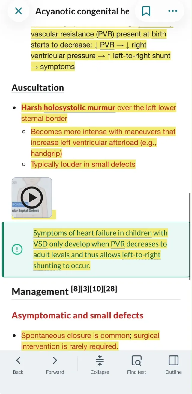
It is most commonly located in the membranous part of the ventricular septum. The defect causes RV volume overload, excessive pulmonary blood flow with pulmonary hypertension, LV volume overload and increased O2 saturation in the right ventricle and pulmonary artery. Symptoms of heart failure in children develop only when PVR decreases to adult levels and allows left-to-right shunting. Auscultation reveals a harsh holosystolic murmur over the left lower sternal border, louder in small defects. Echocardiography is the confirmatory test. Small defects often close spontaneously; large defects require medical management and surgical repair.